
The internal capsule > Lateral view showing corticospinal pathway
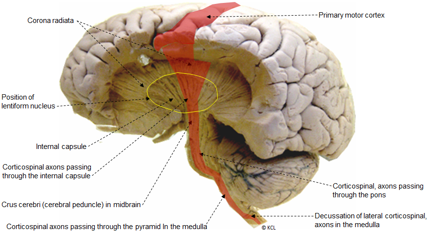
This image shows the internal capsule exposed by dissection, including the position of the corticospinal axons.
The internal capsule (lateral view):
The lentiform nucleus has been dissected away to reveal the curved fan-like shape of the internal capsule. The desending corticospinal tract axons are indicated by the shaded area. Note however that up to 50% of axons forming the corticospinal tract originate from areas adjacent to the motor cortex (premotor cortex and somatosensory cortex), not shown in this illustration.
- Is a band of white matter in the cerebral hemispheres through which almost all tracts enter or leave the cerebral cortex.
- Lies between the lentiform nucleus laterally and the thalamus and head of the caudate nucleus medially.
- In horizontal section is shaped like a shallow letter V, but dissection shows it to be fan-like, with ascending fibres radiating out above it (the corona radiata) towards the different cortical areas, and descending fibres collecting at its base to enter the midbrain.
- Has functionally different tracts that are arranged from anterior to posterior, including somato-sensory, voluntary motor, motor control, vision and hearing as well as those associated with mood, memory and cognitive functions.
- Because of the concentration of different tracts, small lesions to the internal capsule give rise to major functional deficits including paralysis, anaesthesia, loss of motor control, cognitive and memory deficits, etc.