| | |
|||||||
 |
|||||||
| Home > Diagrammatic representations | |||||||
|
|
|||||||
|
|||||||
Copyright (c) KCL 2011 |
|||||||
| | |
|||||||
|
|||||||
| Home > Diagrammatic representations | |||||||
|
|
|||||||
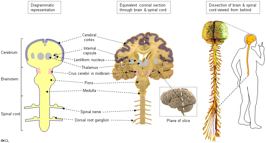
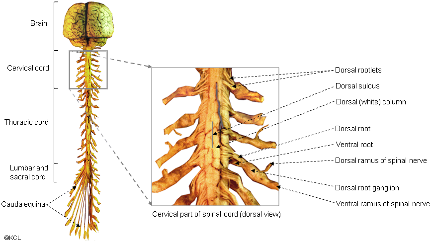
NA1 Explanation of diagrammatic representations: |
|||||||
|
|||||||
Copyright (c) KCL 2011 |
|||||||