Muslce Spindle: Traverse sections through muscle
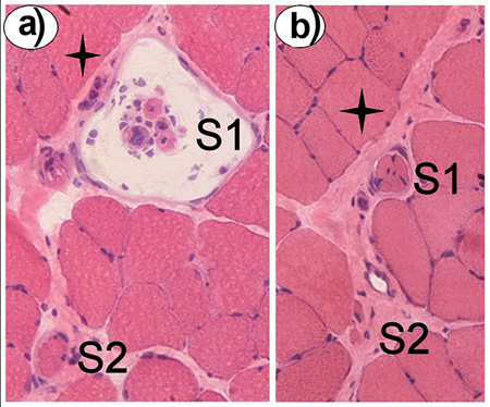
Panels (a) and (b) show transverse sections through muscle. The sections were stained with haematoxylin (purple-blue - for nuclei) and eosin (pinkish-red - for cell cytoplasm and extracellular connective tissue). They are serial sections separated by about 1 mm, and show the same two spindles, S1 and S2, at different positions along their long axis. The same extrafusal fibre is marked with a star in both sections, to help orientation.
In (a), S1 is sectioned through the mid-equatorial region; the intrafusal fibres are grouped together in the middle of the intracapsular space. This is the part which has the afferent nerve terminals, although they don't show up well with the H&E stain. The diameter of the S1 capsule in (a) is about 150 um at this point.
In (b) the same spindle (S1) has caught at the point where the capsule has narrowed down to a thin sleeve around tightly packed intrafusal fibres - this sleeve region is where the motor end-plates are located.
Spindle S2 is already in the sleeve region in (a), and in (b) just the polar ends of the two longest intrafusal fibres (usually the bag fibres) are showing.