Unit 3: Pupil Abnormalities, Facial Nerve Palsy & Ptosis
4: Facial (7th Cranial) Nerve Palsy
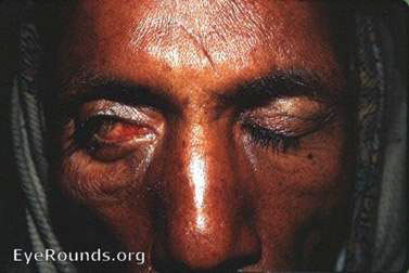
Source: EyeRounds.org Contributor: William Charles Caccamise, Sr, MD, Retired Clinical Assistant Professor of Ophthalmology, University of Rochester School of Medicine and Dentistry.
Causes
- Brainstem disease
- Skull base disease
- Tumours
- Meningeal disease
- Peripheral disease
- Herpes zoster
- Middle ear disease
- Mastoiditis
- Parotid tumours
- Sarcoid (peripheral or meningeal)
- Trauma or surgery
- Idiopathic (Bell's palsy) (over 75%)
Clinical Features
- Paralysis of the facial muscles
- Widened palpebral aperture
- Impaired eyelid closure
- Drooping of the angle of the mouth
Examination
Examine cranial nerves, especially 5th and 6th, and middle ear (otoscopy)
Management
- Consider referral to ENT.
- Refer for neuroimaging if:
- Associated neurological features
- Failure to resolve after 3 months
- Refer to ophthalmology if:
- Corneal exposure
- Epiphora (watering)
- Cosmetic concerns