


- Key concepts
Reticulospinal pathway of descending pain control
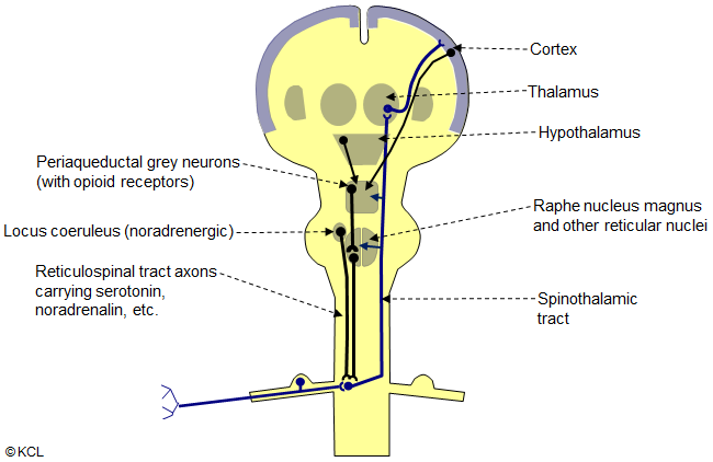
This diagram represents very simply the major features of the descending reticulospinal control of pain, a chain of neurons:
- Neurons in the central midbrain (periaqueductal grey) send axons to the reticular nuclei of the medulla (mainly the nucleus raphe magnus);
- Axons from the reticular nuclei descend in a reticulospinal tract to end in the dorsal horns of the spinal cord. Here they specifically inhibit pain transmission either presynaptically on the terminations of first-order pain fibres in the cord, or postsynaptically on the second order neuron in the dorsal horn via interneurons, so that pain conduction through the spinothalamic tract is blocked.
The neurotransmitters here are serotonin (from the nucleus raphe magnus) and noradrenalin (from some other reticular nuclei).
The periqueductal grey neurons have opioid receptors (targets of opioid analgesic drugs) and are activated by inputs from the cerebral cortex, hypothalamus and other higher brain centres, as well as ascending pain fibres, so that pain transmission can be modulated. In the dorsal horns of the spinal cord, synaptic endings of the reticulospinal axons release serotonin and noradrenalin. Another group of cells in the pons, in the locus coeruleus, send noradrenergic axons into the reticulospinal tract and have a similar inhibitory action on pain transmission in the cord.
Choose a menu item to the left, to continue