Anatomy of the Heart
- Layers
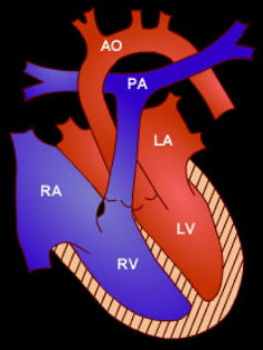
- Chambers
- Valves
- Cardiac Septum
- Cardiac cell & muscle
- Conduction system

Move your mouse over the images to see a larger version.
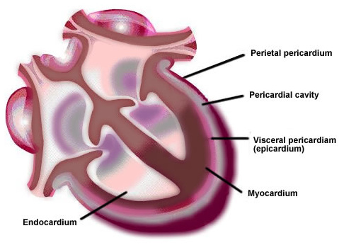
The wall of the heart is composed of three distinct layers:
- Epicardium (outermost layer of the heart)
- Myocardium (intermediate layer)
- Endocardium (inner lining of the cardiac chamber)
Move your mouse over the images to see a larger version.
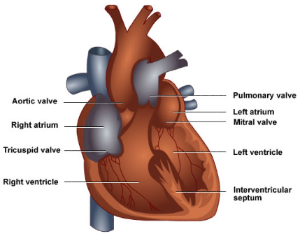
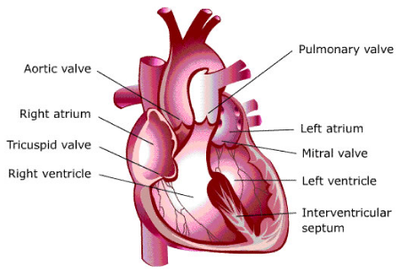
The heart contains four valves:
The interatrial septum (which is difficult to visualise on the image) separates the two atria.
The interventricular septum (labelled) divides the ventricles.
Separating the atria above and the ventricles below is the atrioventricular septum.
The cardiac cell contains bundles of protein strands called myofibrils. Myofibrils are surrounded by sarcoplasmic reticulum, which contains cysternae. The sarcomeres are the contractile unit of myofibrils.
Cardiac muscle is an involuntary striated muscle, which is mononucleated. Cardiac muscle has cross striations formed by alternation segments of thick and thin protein filaments (anchored by segments called Z-lines). Cardiac muscle is relatively shorter than skeletal muscle.
Actin and myosin are the primary structural proteins. Under light microscope, the thinner actin filaments appear as lighter bands, while thicker myosin filaments appears as darker bands. The thinner actin flaments contain two others protiens called troponin and tropomyosin which plays a important role in contration.
Cardiac muscle also contains dense bands (specialised cell junctions) called intercalated discs that separate individual cells from one another at their ends.
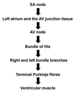
Image: Sequence of cardiac excitation
The cardiac conduction system consists of highly specialised cells that histologically resemble nerve tissue.
These are important in maintaining the heart's electrical activity in an orderly fashion.
Five distinct anatomical structures comprise the cardiac conduction system:
- Sinoatrial (SA)/sinus node
- Atrioventricular (AV) node/node of Tawara
- AV bundle of His
- Right and left bundle branches
- Purkinje fibers