Physiology of the Heart
- Contraction and relaxation
- Circulatory system
- Circulatory pathway
- The cardiac cycle
- Heart sounds
- Mechanism of contraction
The contraction and relaxation of the heart involves both:
1] Systole: This is the period of contraction during which the ventricles or atria force blood from chamber to chamber and through the blood vessels.
2] Diastole: This is the period of relaxation during which the ventricle or atria fill with blood.
The blood flow in the coronary arteries is maximal during diastole and minimal in systole.
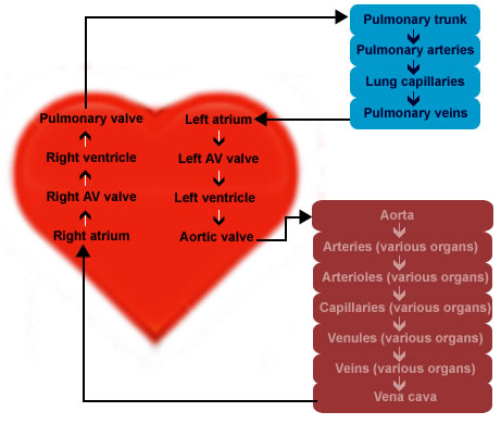
The diagram shows the circulatory pathway throughout the cardiovascular system. Blood flows from the left ventricle to the arteries, arterioles, capillaries of the body, it then flows to the venules, veins, right atrium, right ventricle, pulmonary artery, lung capillaries, pulmonary veins, left atrium and finally back to the left ventricle.
Move your mouse over the images to see a larger version.
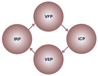
Phases of cardiac cycle
The first phase of the cardiac cycle is the VFP which occurs during diastole. At the beginning of the VFP, the heart is in a polarised state and blood is moving through the atria into the ventricles.
As an electrical stimulus occurs from the S-A node, atrial cells depolarise and the atria subsequently contracts. This contraction forces additional blood past the tricuspid and bicuspid/mitral valves, filling the ventricles.
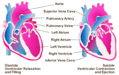
Shows the ventricular relaxation and
filling phase (diastole)
and ventricular contraction
and ejection (systole) phase of the cardiac cycle
The cardiac cycle is the sequence of events that occur when the heart beats. It consists of cyclic structural and functional changes that the heart undergoes every 0.8 of a second (on average) to maintain the blood flow through the body. The cardiac cycle consists of contraction and relaxation of both atria and ventricles. The cardiac cycle is divided into four separate periods.
Click on each phase for more information
The second phase of the cardiac cycle is the ICP which begins systole for the cardiac cycle which is the beginning of ventricular contraction.
During this phase the blood is not being ejected from the ventricles, but the pressure is building in the ventricles in order to force aortic and pulmonary valves to open. The pressure in the ventricle must exceed the pressure in the aorta for blood to be ejected from the heart.
The third phase of the cardiac cycle is the VEP which is a continuation of the systolic phase of the cardiac cycle. At this stage the pressure within the ventricles has increased well above the pressure in the aorta and pulmonary vein and the pressure difference forces the semilunar valves to open and blood is ejected from the ventricles into the arteries.
Blood will flow past the aortic and pulmonary valves until the pressure gradient in the arteries exceeds the pressure of the contracting ventricles. Upon equilibrium of the pressures between the ventricles and arteries, the aortic and pulmonary valves will shut, and blood flow from the ventricles will cease.
Two of the periods (VFP and IRP) occur during the diastole (relaxation phase) and two periods (ICP and VEP) occur during the systole (contraction phase).
The first heart sound (S1) occurs with the closing of the mitral and tricuspid valves at the beginning of ventricular systole.
The second heart sound (S2) occurs with the closing of the aortic and pulmonary valves at the onset of ventricular diastole.
Systole is the contraction of the heart that drives blood into the pulmonary arteries and the aorta. The first heart sound (S1) indicates the onset of systole.
Diastole is the period between contractions of the atria and ventricles (blood enters the relaxed chambers of the heart). The onset of the second heart sound (S2) is the beginning of ventricular diastole, and the diastole ends at the onset of the first heart sound (S1).
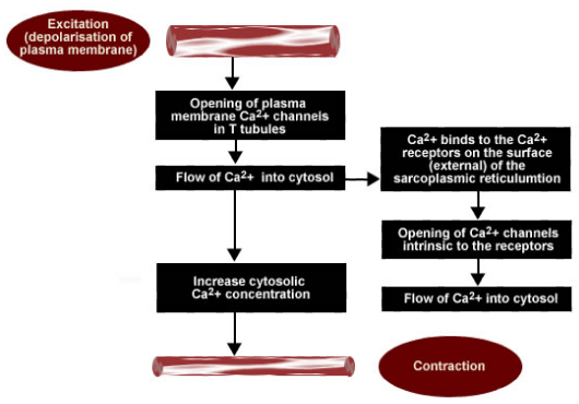
Shows the contraction mechanism of the heart
Move your mouse over the images to see a larger version.
The regulation of cardiac muscle contraction is carried out by neural, hormonal and intrinsic components. The heart is composed of cylindrical cardiac cells and stimulation of one cardiac cell initiates stimulation of adjacent cells and finally leads to cell contraction.
In general, the cardiac cells are of two types, electrical cells and myocardial cells. The myocardial cells have two specific properties such as contractility (ability of the cells to shorten and return to their original length) and extensibility (cell filaments ability to stretch).
The contraction mechanism contains numerous steps and excitation and contraction coupling is the term used to define these events, which connect the depolarisation of the cardiac cell membrane to the contraction of the muscle fibers.