Coronary Circulation
- Arterial circulation
- Venous circulation
- Coronary Collaterals
- Cardiac Lymphatics
- Nerve supply of heart
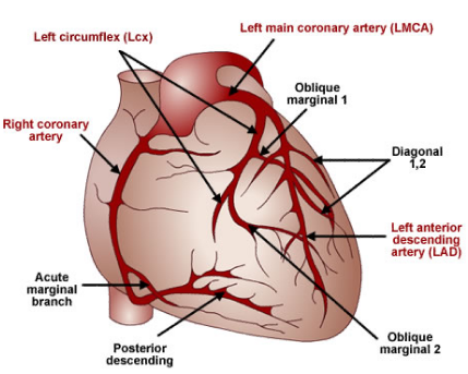
Coronary arteries supply blood to specific areas of the heart.
Move your mouse over the images to see a larger version.
The coronary arteries supply blood, oxygen, and nutrients to the heart muscle and the conduction system.
The two principal coronary arteries are:
- Left main coronary artery (LMCA)
- Right coronary artery (RCA)
The left main stem bifurcates to form the:
- Left anterior descending artery (LAD)
- Left circumflex artery (LCx)
Click on the title below for more information.
The right coronary artery supplies blood to:
- Right venticle
- Right atrium
- Diaphragmatic surface/inferior wall of LV
- Posterior wall of left ventricle (90%)
- Posterior third of interventricular septum
- SA node (55-60%)
The LMCA/LAD supplies blood to:
- Anterior and lateral wall of LV
- Most of the left ventricle
- Interventricular septum (anterior 2/3rd)
- Right and left bundle branches
The LMCA/LCx supplies blood to:
- SA node (40-45%)
- Left atrium
- AV node and bundle of His (10%)
- Lateral wall of LV
The coronary collaterals is a channel created to bypass an obstructed blood pathway by enlarging secondary blood vessels.
It also provides communication between major coronary arteries and branches.
It may dilate and provide blood supply beyond the obstructed/stenosed epicardial vessel.
Lymphatics drain towards the epicardial surface and they merge to form the right and left channels.
Left and right channels travel in retrograde fashion with their respective coronary arteries.
The left and right channels travel along the ascending aorta and merge before draining into pretracheal lymph node.
The merged single lymphatic chain travels through a cardiac lymph node and finally empties into the right lymphatic duct.
Coronary arteries supply blood to specific areas of the heart.
Move your mouse over the images to see a larger version.
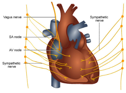
The sympathetic and parasympathetic send fibres to the heart, which form the cardiac plexus.
The cardiac plexus is located close to the arch of the aorta.
The parasympathetic vagus fibres act as inhibitors and reduce the heart rate and stroke volume.
The sympathetic nerves act as accelatory nerves accelerating the heart rate and increasing the stroke volume.

