This technique is used to evalute left ventricular function during routine myocardial perfusion study. The gating principle is the same. Different software is used in routine practice.
Software allows endocardial and epicardial border recognition. The area within a cardiac slice is calculated as the area inside the endocardial border. The left ventricular volume is calculated by adding up the areas of each cardiac slice e.g. if the total left ventricular cavity is divided into 8 slices, the volume would be the sum of the areas of the 8 slices.
Click on the Examples tab above to continue.
The following images show the calculation of LV volume and ejection fraction.
Click on each title for more information.
Image 1
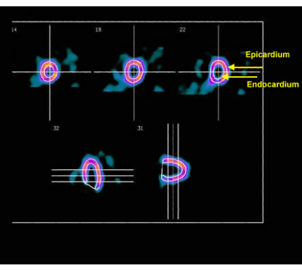 Shows the endocardium and epicardial contours (arrows).
Image 2
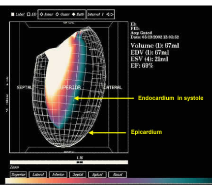 Shows the outline of the LV in 3D in systole. The colour scale shown at the bottom of the image shows the relative uptake of tracer by the different left ventricular walls where white represents maximum uptake and aqua-marine represents minimum uptake.
Image 3
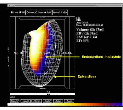 Shows the outline of the LV in 3D in diastole. On this and the next image, white represents maximum uptake and blue represents minimum uptake.
Image 4
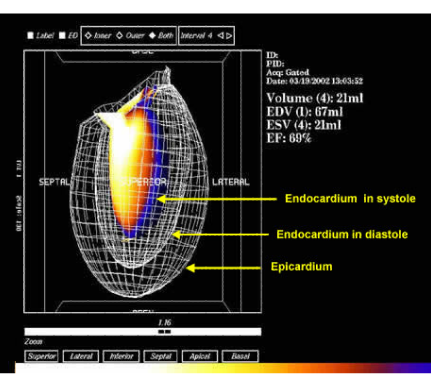 The area within each left venticular slice is added up to derive the final left venticular volume. This is done in both
diastole and systole. The image shows the outline of the LV epicardium and endocardium in 3D
The area within each left venticular slice is added up to derive the final left venticular volume. This is done in both
diastole and systole. The image shows the outline of the LV epicardium and endocardium in 3D
Image 5
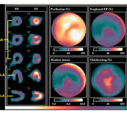 Shows the representative LV slices in diastole (ED) and systole (ES) in three different orientations; short-axis (SA), vertical long-axis (VLA) and horizontal long-axis (HLA) on the left hand side. The polar plots are demonstrated on the right hand side are a semi-quantitative estimate of myocardial perfusion, regional ejection fraction, wall motion and wall thickening
Image 6
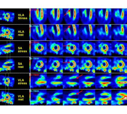 This example of a myocardial perfusion scan. In the image, there is a comparison made between stress and rest images as shown. The red arrows point to a fixed defect in the antero-septal wall and apex in keeping with a large MI.
Image 7
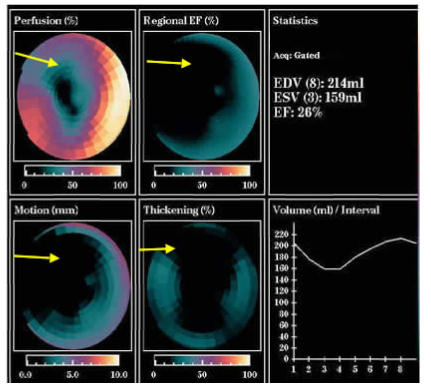 This is the corresponding GSPECT image showing the reduced perfusion, wall motion and thickening in the infarcted
area (shown by the arrows). The LVEF is depressed = 26%
This is the corresponding GSPECT image showing the reduced perfusion, wall motion and thickening in the infarcted
area (shown by the arrows). The LVEF is depressed = 26%
Click the Next button below to continue.