2] Enteral devices
A number of tubes are inserted into the enteral cavity for feeding and monitoring.
Nasogastric Tube (NG Tube)
An NG feeding tube is a long plastic tube used to allow feeding of a patient unable to take water or food orally. It passes from the nostril to the stomach and should follow the line of the oesophagus and pass below the level of the diaphragm. It is seen as a faint linear density on the X-ray. On placement of an NG tube into the patient, the position of the tip in the stomach, is usually confirmed by aspirating a few mls of gastric content and testing on litmus paper for acidity. An X-ray is only performed if this method is inconclusive or reveals alkaline contents.
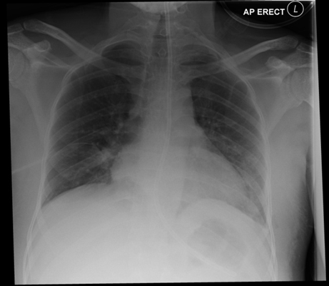
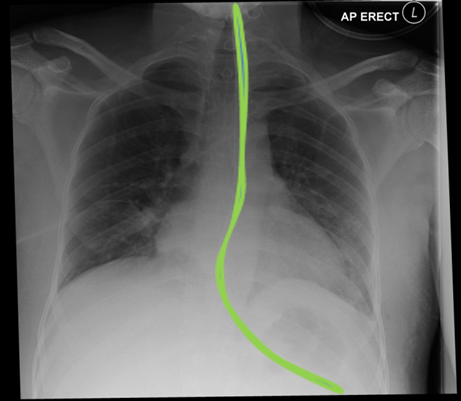
Here is a Chest X-ray showing correct placement of a Nasogastric tube with the tip in the stomach.
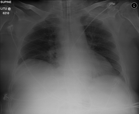
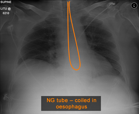
This patient has had an NG tube sited but the tip has double backed on itself and therefore would not be functional as a means of inserting food/ fluid into the stomach. The tube needs to be re-sited before it can be used safely. The tip should lie over the stomach bubble seen as a dark bubble under the left hemidiaphragm.
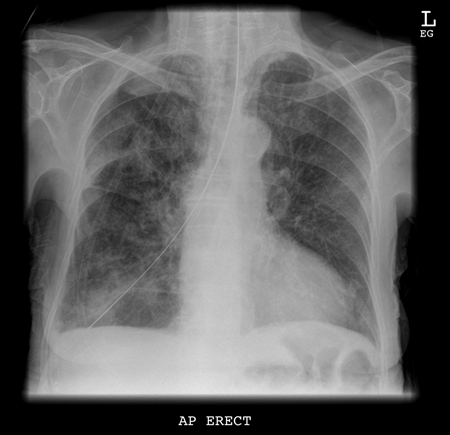
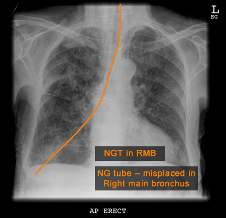
This patient has had an NG tube inserted and the tube has passed down the trachea, right main bronchus (RMB) and into the right lower lobe. If this patient was to be fed through the tube in this position, the patient would develop an aspiration pneumonia and could potentially die. This tube needs to be re-sited with the line following the direction of the oesophagus with the tip in the stomach before it can be used safely.
Impedance catheter
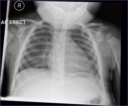
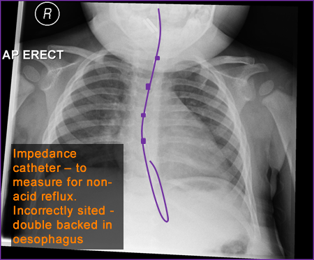
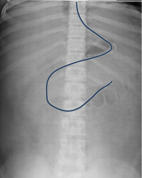
Another type of line also seen in the same position as an NG (nasogastric) tube is an impedance catheter (purple tube). It looks similar but has sensors seen along the length of the tube. This is inserted temporarily as an investigation that measures pressure differences in the oesophagus which relates to non-acid reflux.
This particular impedance catheter has been inserted incorrectly and the tip is double backed in the oesophagus instead of lying in the stomach.
Nasojejunal tube
For long term feeding, a nasojejunal tube is sometimes used where the tip is inserted further along towards the jejunum. This is especially needed if the patient suffers with reflux of gastric contents up the oesophagus.
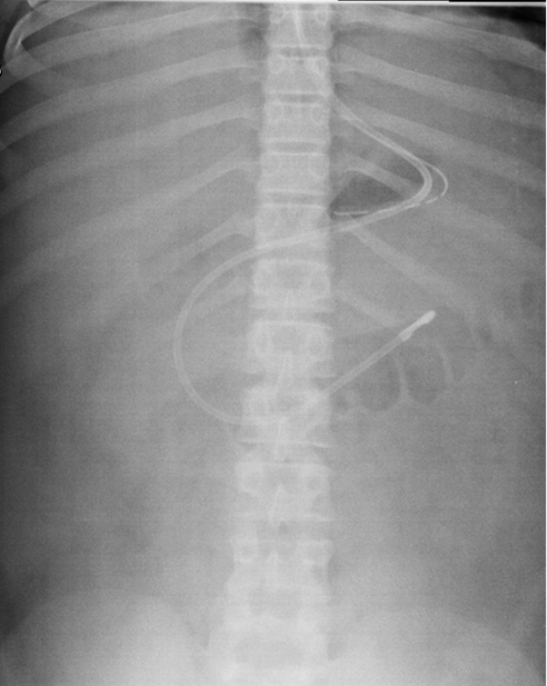
Here is an example of a nasojejunal tube which has been placed into the patient. It is differentiated from the NG tube as it often has a radio-dense (white) tip. It is passed via the nostril down the oesophagus and into the stomach. It is further advanced out of the stomach and follows the line of the duodenum until the tip lies at the duodenojejunal junction which should lie to the left of the spine. It can sometimes be passed under fluorsoscopic (Xray) guidance in the radiology department or via endoscopy (camera) if not successful on the ward.
Gastrostomy tube
Again for long term feeding , a gastrostomy can be made surgically for direct access into the stomach.
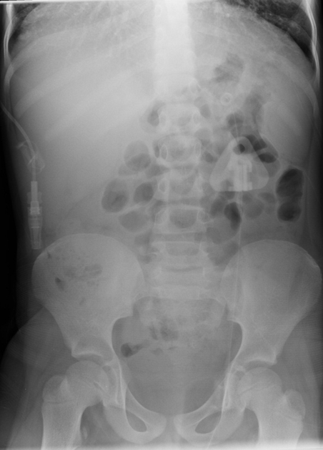
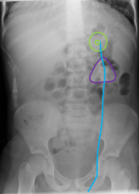
Here is a patient who has had a PEG or Percutaneous Endoscopic gastrostomy inserted.It is a tube which is inderted directly into the stomach via the skin either surgically or radiologically (under Xray guidance) to allow feeding directly into the stomach if there are issues regarding long term safety of feeding. There in an internal retention disc (green) inside the stomach, an external tube (blue) and clamp and external fixation plate (purple).
