3] Ventilatory devices
A number of tubes are inserted into the enteral cavity for feeding and monitoring.
Endotracheal Tube (ET tube)
An ET tube is a short plastic tube used to ventilate a patient artificially such as in an operating theatre or in intensive care. It is inserted via the mouth into the trachea (Link to airway anatomy of trachea and bronchi).
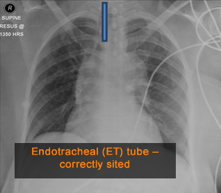
It can be identified on a chest X-ray as two parallel lines one line more dense (whiter) than the other. It should lie in the midline overlying the trachea with the distal end midway between the thoracic inlet and the carina. The tip should lie approximately 2 cm above the carina (1cm in children).
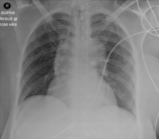
Here is a patient’s CXR that shows an endotracheal tube in place.
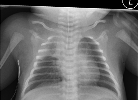
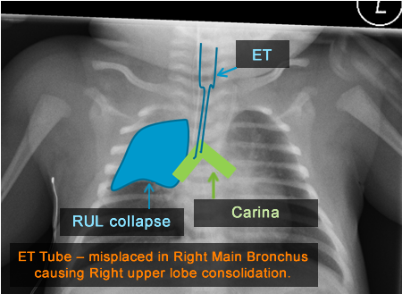
In this patient, the ET tube lies too far and is within the right main bronchus, near the bronchus intermedius. The right upper lobe is therefore not receiving adequate ventilation and therefore is looking more dense (white) than it should, and is starting to collapse.
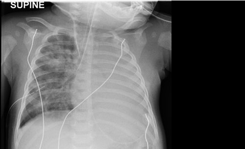
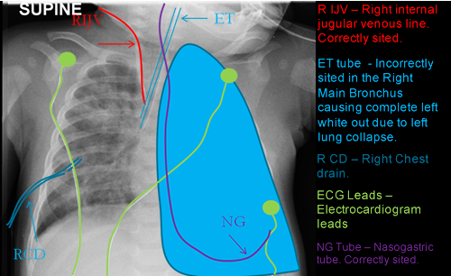
In this patient, the ET tube is again sited in the right main bronchus and there is no adequate ventilation reaching the left lung which looks of increased density (blue shaded area). A chest drain (RCD)is also present in the right chest cavity draining a previous effusion or pneumothorax (fluid or air in the pleural space).
A right internal jugular vein central venous catheter (RIJV) is in place for venous access. The tip is correctly sited in the superior vena cava.
An NG tube is correctly sited with the tip over the stomach bubble. ECG leads ( green leads) are also present on the chest wall.
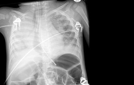
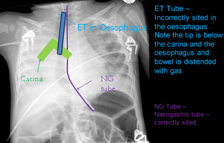
The ET (endotracheal tube) is seen to pass below the level of the carina (green shaded) and therefore it cannot lie in the trachea. Also of note, the oesophagus and bowel below the diaphragm is distended with gas (looks blacker). These signs should alert us that the tube may not be in the trachea. The right lung is also not adequately aerated. In this case the ET tube is sited in the oesophagus and needs to be changed.
The NG tube is adequately sited.
Tracheostomy tube and Surgical Artefacts
In chronically ventilated patients the endotracheal tube is often exchanged for a tracheostomy tube. A tracheostomy is first performed which creates a hole in the anterior aspect of the neck directly to access the trachea and the tube is then inserted directly into the trachea.
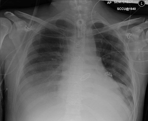
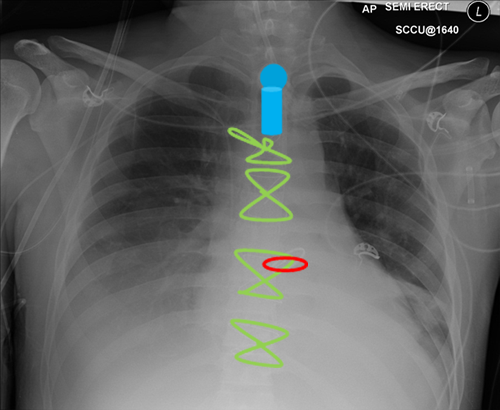
This patient has a tracheostomy tube in place (Blue shaded area).
In addition, the patient has Sternotomy clips (Green) which are as a result of cardiac surgery involving opening the chest cavity.
This patient has had a Mitral valve replacement (Red). There are also ECG leads seen on the chest wall.
