4] Intravascular devices
Very sick patients in intensive care environments or chronically ill patients often have tubes and devices inserted into their vessels. This provides temporary or long term access to the vessels to allow administration of a number of items including intravenous medication, nutrition, long term antibiotics or chemotherapy as well as allow haemodialysis, blood sampling and monitoring or protection of the patient. Vascular access can be central such as an Internal jugular, subclavian line or portacath or peripheral such as PICC lines. They are inserted through the skin and the tip should lie in a central vessel, near the heart. Some other common devices seen include ECMO catheters, Swan-Ganz catheters and IVC filters.
Internal Jugular Vein (IJV) catheter
An internal jugular venous plastic catheter tube which is inserted into a central major vein in the neck (internal jugular vein), allowing rapid access to the venous system for fluid and drugs. It usually lies on the right side and should follow the line of the internal jugular vein and the tip should lie near the Superior vena cava. A chest X-ray is taken after placement of the line to confirm the position of the tip and also look for complications such as a pneumothorax (puncture of the lung with air in the pleural space).
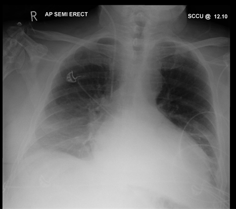
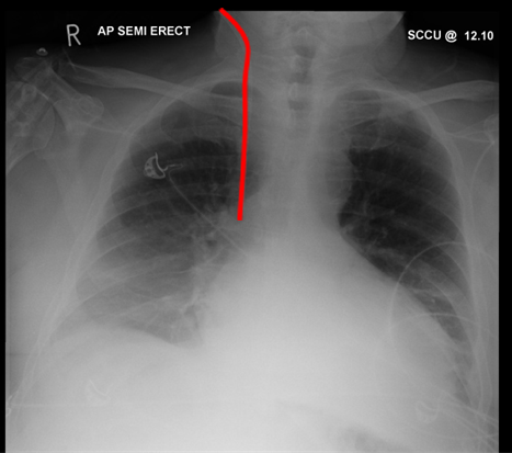
This patient’s Chest X-ray shows ECG leads and a right Internal Jugular line. There are no complications of placement such as a pneumothorax.
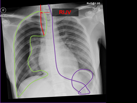
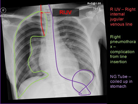
This Chest X-ray shows a patient who has had a right internal jugular line (RIJV) placed which has caused a pneumothorax (green area) as a complication. The patient also has a coiled up Ng tube in the stomach (purple line).
Subclavian venous (SCV) catheter
A subclavian venous catheter is a similar catheter tube which is inserted into the subclavian vein and again the tip should lie at the superior vena cava. The risk of a pneumothorax or haemothorax is slightly higher for this type of catheter than an internal jugular one.
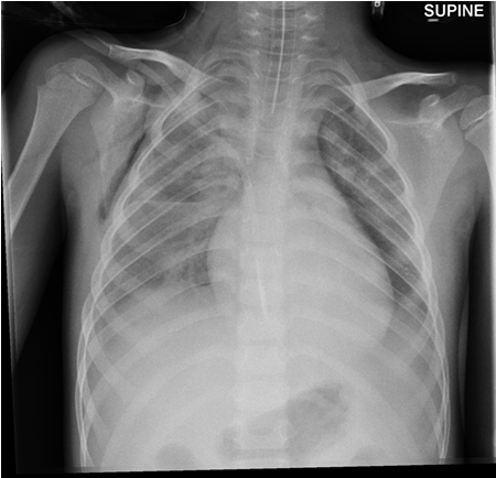
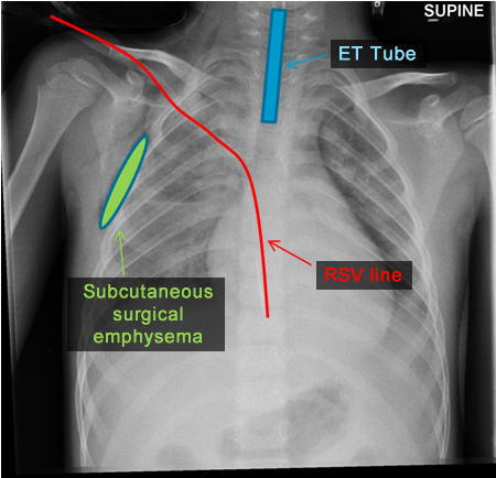
The right Subclavian venous line tip in this patient lies too far within the heart and lies in the right atrium instead of the SVC. As the line tips are thrombogenic, if left in the atrium there is increased risk of a thromboembolism (blood clots to form and pass through the circulation) to occur. This patient is also noted to have air within the subcutaneous tissues of the right chest wall known as subcutaneous emphysema.
Portacath
A portacath is another form of vascular access often used for long term venous access often in an outpatient setting. It is often seen in patients with cystic fibrosis or leukaemia and can be used to take regular blood samples or for administration of chemotherapy or long term antibiotics. The port lies on the chest wall and tubing is tunnelled along the chest wall and enters the central venous system at the brachiocephalic veins. The tip should lie at the superior vena cava.
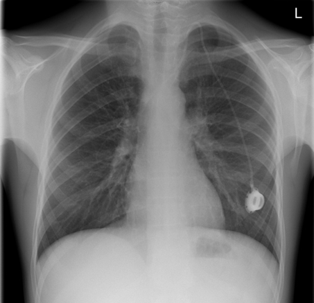
This patient has a Portacath fitted for intravenous access seen on the Chest X-ray. The tip is at the Superior Vena Cava.
PICC Line (Peripherally inserted Central Catheter)
A PICC line is another type of intravenous line usually inserted by an interventional radiologist. It is placed usually in the arm into the brachial vein and the tip should lie centrally at the SVC. It can stay in place as long as the intravenous therapy or nutrition is required.
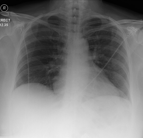
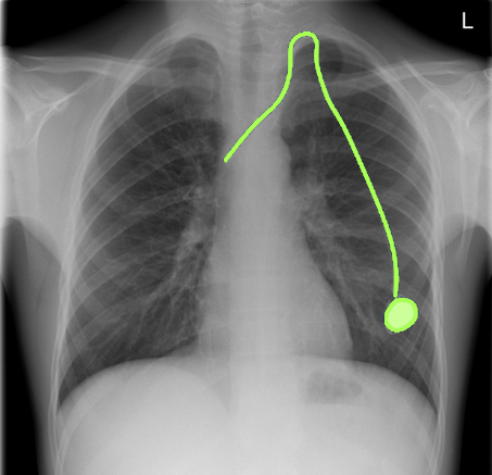
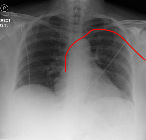
This patient has a left sided PICC line (red) in situ. It has been inserted into the left brachial vein and the tip of the PICC line is seen in the SVC/Right atrial junction.
When assessing X-rays for lines and tubes, it is not only important to look for the tubes positions but also to look for complications of there placement. Complications such as pneumothoraces ( air in the pleural space) or haemorrhage or retained guide wires are some examples.
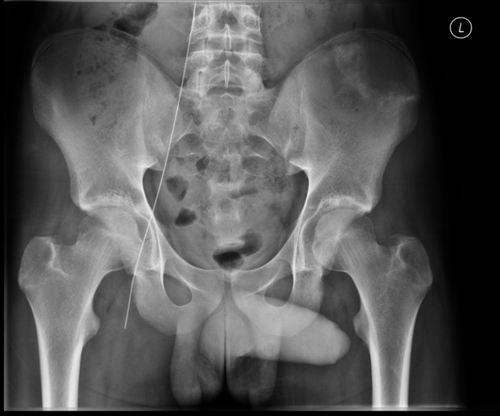
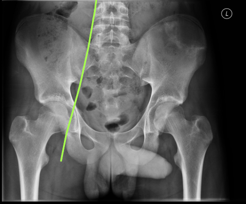
This patient had a retained guide-wire after a femoral venous line was inserted into the groin for venous access. It had to be retrieved by the interventional radiology department on discovery of the wire.
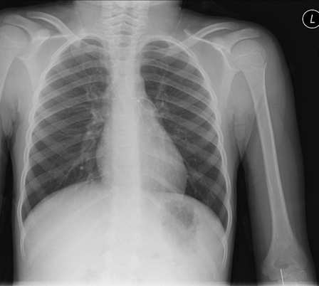
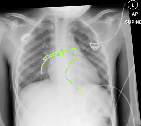
This patient had a PICC line inserted in the left antecubital fossa. Unfortunately it snapped and was seen a day later coiled inside the pulmonary trunk and then later seen in the right and left pulmonary artery (blue line). It had to be removed by cardiothoracic surgeons although often these can be removed by interventional radiologists. If left the patient is at risk of a pulmonary embolism (clot) within the pulmonary arteries.
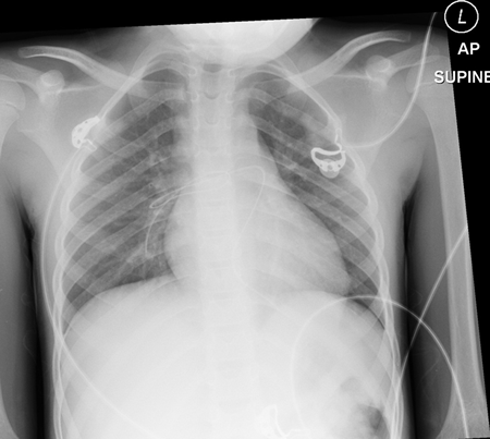
This patient had a PICC line inserted in the left antecubital fossa. Unfortunately it snapped and was seen a day later coiled inside the pulmonary trunk and right and left pulmonary artery (blue line). It had to be removed by cardiothoracic surgeons although often these can be removed by interventional radiologists. If left the patient is at risk of a pulmonary embolism (clot) within the pulmonary arteries.
Extracorporeal Membrane oxygenation (ECMO)
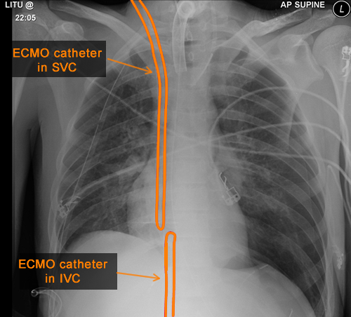
This form of support for the patient is used when conventional methods of circulatory and ventilator support have failed. It can involve V-V (veno-venous) or V-A (veno-arterial) exchange of blood and requires tubes being placed in the Superior Vena cava and Inferior vena cava or brachiocephalic artery.
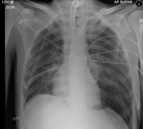
This patient has ECMO support and has veno-venous exchange with a catheter in the Superior vena cava and a further catheter in the inferior vena cava (orange tubes). The patient also has a tracheostomy, an NG tube, a right sided chest drain and ECG leads on the chest.
Swan-Ganz catheter
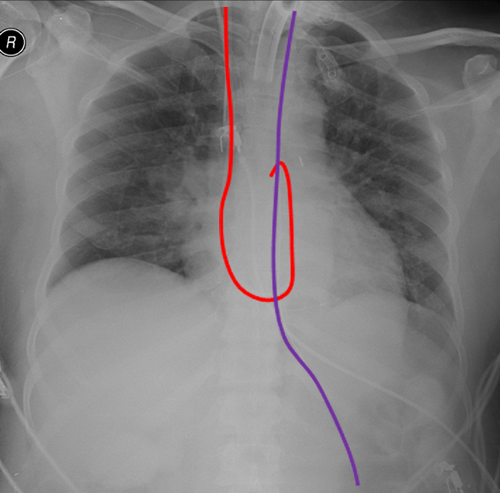
The Swan-Ganz catheter is an intravascular tube inserted into the right side of the heart with the tip in the pulmonary arteries. It is used in sick patients in an intensive care facility to measure and monitor right sided heart pressures. It can be seen on a Chest-Xray as a catheter forming a loop projected over the heart.
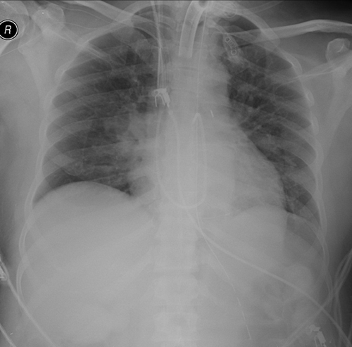
This patient has a Swan-Ganz catheter in place seen extending from the right internal jugular vein in the neck through the SVC, right atrium and ventricle and into the pulmonary artery trunk (Red Line). The patient also has a tracheostomy tube in place to help with chronic ventilation, an NG tube (Purple line), a right sided IJV line and ECG leads.
Inferior Vena cava filter
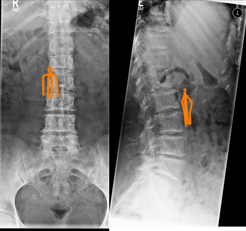
An IVC filter is an umbrella shaped medical device that acts as a vascular filter that is placed in the Inferior vena cava in bed bound patients. It prevents blood clots getting to the lungs and causing blockages of the pulmonary artery vessels (pulmonary emboli). They are placed by interventional radiologists into the IVC from the groin.
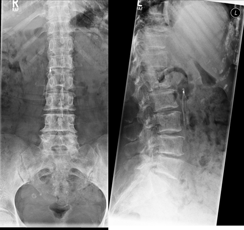
This patient has an IVC filter in place. It can be seen as a metallic umbrella shaped device (orange) which is placed in the Inferior Vena Cava to catch clots / thrombi and stop them reaching the lungs in bed bound patients.
