5] Intracavitary devices
Some lines and tubes need to be placed inside a cavity of the body to perform its function. They are mostly inserted into the pleural or peritoneal cavity.
Chest drain
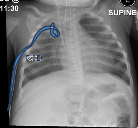
When a patient in hospital develops either a Pneumothorax or Pleural effusion (links to previous) the treatment involves placement of a chest drain. This is a plastic tube which has an end hole and multiple side holes at the distal tip. When inserted into the chest cavity via the skin in between the ribs can allow drainage of the air or fluid and allow the lung to re-expand. This is done as a procedure by trained staff with local or general anaesthetic. An X-ray is then performed to confirm the position of the tip of the drain to lie within the fluid or air pocket. The tip is usually placed in the apex for pneumothoraces and at the base, posteriorly for pleural effusions.
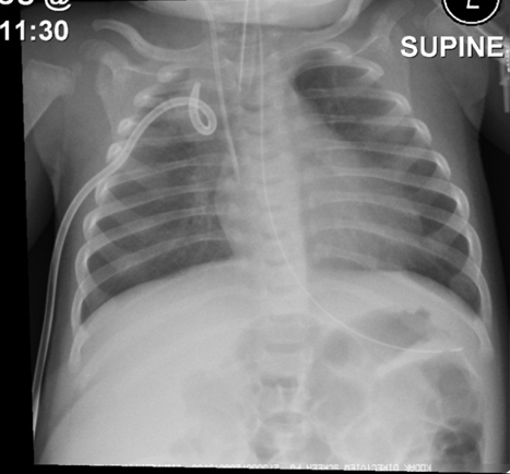
This child on the paediatric intensive care unit has had a right sided chest drain inserted for a previous pleural effusion which has now been drained away. It has a pigtail end.
Ventriculoperitoneal Shunt
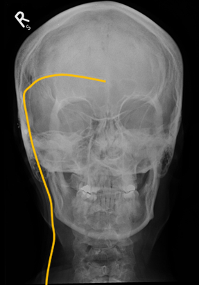
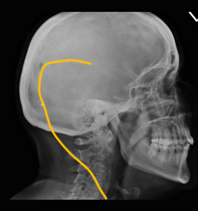
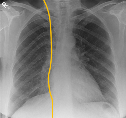
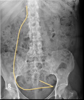
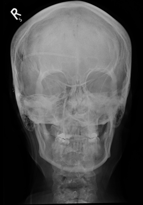
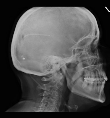
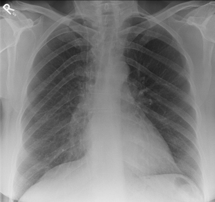
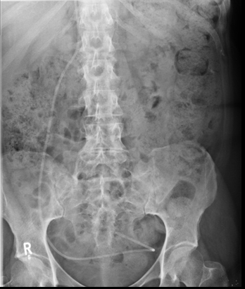
A further catheter that is used in patients with raised intracranial pressure is a VP shunt (orange line). This involves an operation to place a catheter from the intracranial ventricles to the peritoneum. This is to drain excess cerebrospinal fluid from the ventricles into the peritoneal cavity. To image the shunt a number of radiographs need to be performed in a Shunt series.
This patient has got a Ventriculo-peritoneal shunt ( orange) in place to drain excess cerebrospinal fluid. A Shunt series has been performed to check for continuity of the V-P Shunt looking for breakages in the tubing.
Peritoneal Dialysis Catheter
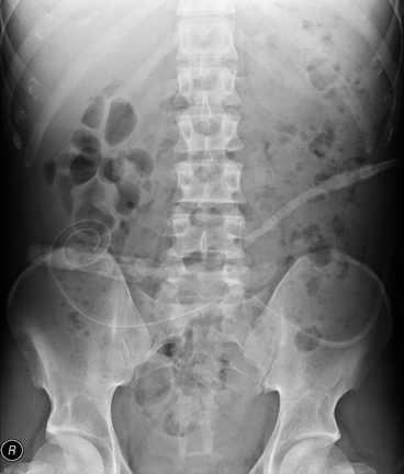
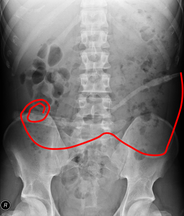
This patient has kidney failure and requires dialysis to remove toxins from the blood. Conventional haemodialysis requires a patient to be attached to a machine for several hours whilst the blood is removed, cleaned and returned to the body. Another form of dialysis is ambulatory peritoneal dialysis which allows patients to be more mobile. The peritoneal catheter lies in the peritoneal cavity (Red).
