MRI clinical applications
- Functional neuroimaging

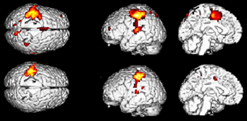
Functional images of the brain from the Institute of Psychiatry, KCL
- If a patient is presented with sensory information of asked to perform a motor task, the cortical region responsible for this activity receives an overabundant supply of oxygenated blood.
- The magnetic properties of oxygenated and deoxygenated blood differ, so if the patient is imaged in a resting state and performing the task then the signal intensity in the relevant cortical region will differ slightly (a few percent at most) in the two cases.
- Computer analysis allows this difference to be displayed as an activation image, as above.
- This technique has applications in basic neuroscience, and in planning neurosurgery for individual patients so as to preserve as much neurological function as possible.