Unit 1: Sudden Loss of Vision
3: Acute angle-closure glaucoma
Anatomy

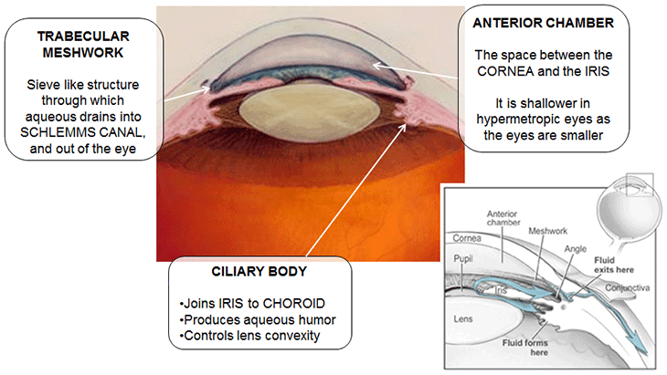
Anatomy of the eye
Source: Main image adapted from CC licensed 'Drawing of the Eye', National Eye Institute, National Institutes of Health (cropped and rotated, labelled, and overlaid with 'Fluid flow', National Eye Institute, National Institutes of Health).

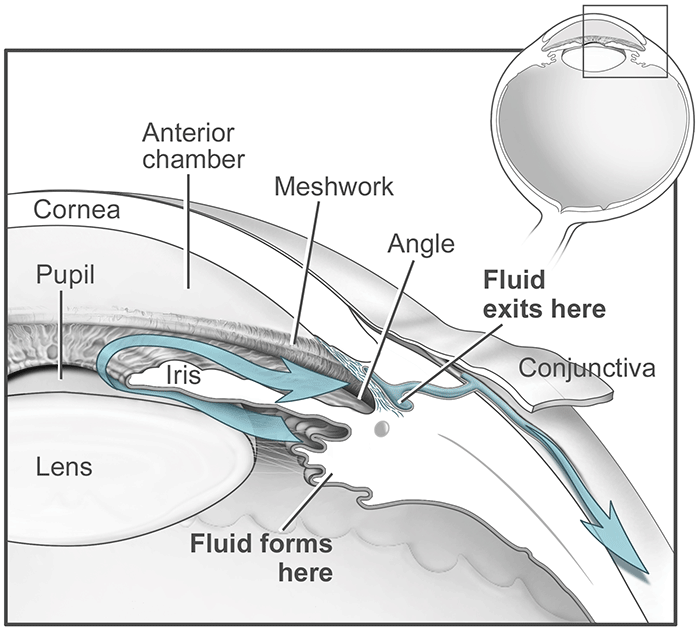
Anatomy of the eye showing fluid flow
Source: Drawing of the Eye. Credit: National Eye Institute, National Institutes of Health. CC-BY-2.0
Physiology
Aqueous production and drainage is balanced to maintain an appropriate intraocular pressure. Aqueous humor is produced by the ciliary body in the posterior chamber. It circulates to the anterior chamber, through the pupil, and leaves the eye through the trabecular meshwork.