Opthalmology
Learning Units
Unit 1: Sudden Loss of Vision
Unit 2: Visual field defects, double vision & optic disc swelling
Unit 3: Pupil abnormalities, Facial nerve palsy & Ptosis
Unit 4: Refractive Error
Unit 5: Children & Squint
Unit 6: Differential diagnosis of blurred vision
Unit 7: Gradual Loss of Vision
Unit 8: Eye Trauma
Unit 9: Red Eye
Unit 10: Systemic Disease
Useful Links
KEATS
KCL website
Contact Us
Unit 1: Sudden Loss of Vision
Introduction
1: Non-arteritic anterior ischemic optic neuropathy
Risk Factors
History & Exam
Symptoms
Signs
Management & Prognosis
2: Giant cell arteritis
Intro
Symptoms
Signs
Examination
Management
3: Acute angle-closure glaucoma
Anatomy & Physiology
Mechanism
Risk Factors
Symptoms
Signs
Treatment
4: Retinal detachment
Anatomy & Physiology
Retinal Tears
Risk Factors
Symptoms
Examination
Management
Self Assessment
1: Non-arteritic anterior ischemic optic neuropathy
Management
Refer to Ophthalmology urgently, to exclude
giant cell arteritis
(link opens in a new Window/tab) with examination /ESR/CRP
Review and treat all cardiovascular risk factors
Consider aspirin
Prognosis
30% make substantial improvement (gain of 3 lines on a Snellen vision chart)
15-50% risk to fellow eye
The optic disc swelling gradually resolves, leaving optic disc pallor (due to loss of neural tissue)
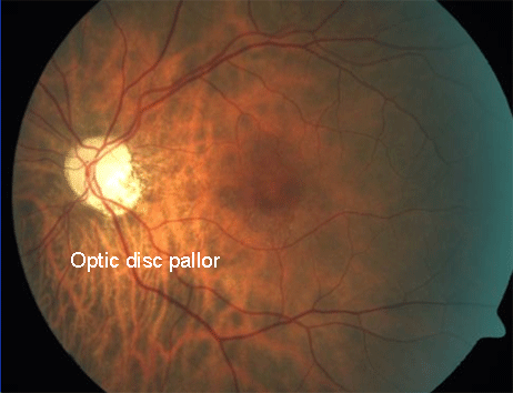
Optic disc pallor
Source: www.retinagallery.com
«
Top
»