2] Radiographic Views
There are several different views that can be taken of the chest which can alter the appearance of the CXR and therefore the interpretation. These are named by the direction the beam through the patient.
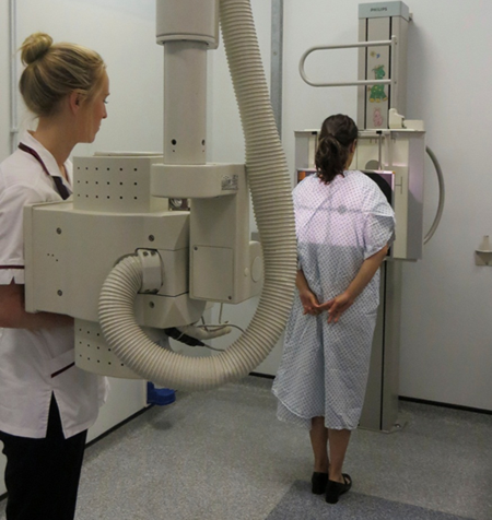
PA – Back to Front (Department)
The "gold standard" is the PA (postero-anterior) CXR taken in all Radiology departments. This is perfect for well adult patients and older children walking into the department.
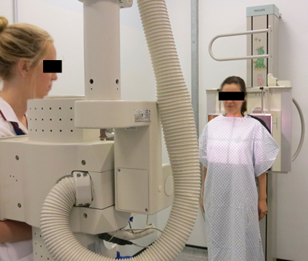
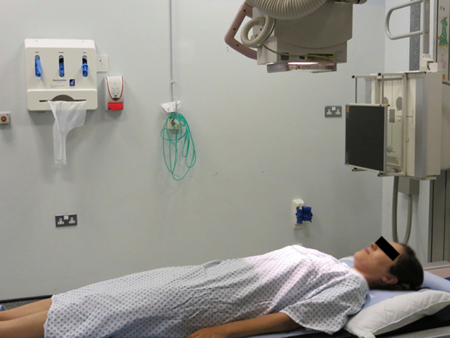
AP – Front to Back ( ITU)
For those that are unwell in Intensive care units, the AP (anteroposterior) view has to be performed and causes problems with interpretation of heart size (described later).
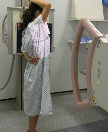
Lateral
Lateral views are used less and less nowadays but can help with localisation of pathology.
Supine ( ITU, Babies)
Supine views are used again in intensive care settings when patients are on ventilators for breathing and also for babies.
Variation of Appearances with Different Views
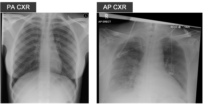
The heart appears bigger on an AP film making it difficult to assess for heart size.
The reason why the heart size varies between AP and PA views is due to cardiac magnification from a divergent X-ray beam. In a PA view, the heart sits anteriorly against the anterior chest wall and therefore as the beam passes through the edge of the heart there is less time for the beam to diverge before hitting the film. In an AP film, the beam goes through the edge of the heart and diverges further before hitting the film. The heart therefore appears much larger on the screen than it should do. The chest wall size does not change.
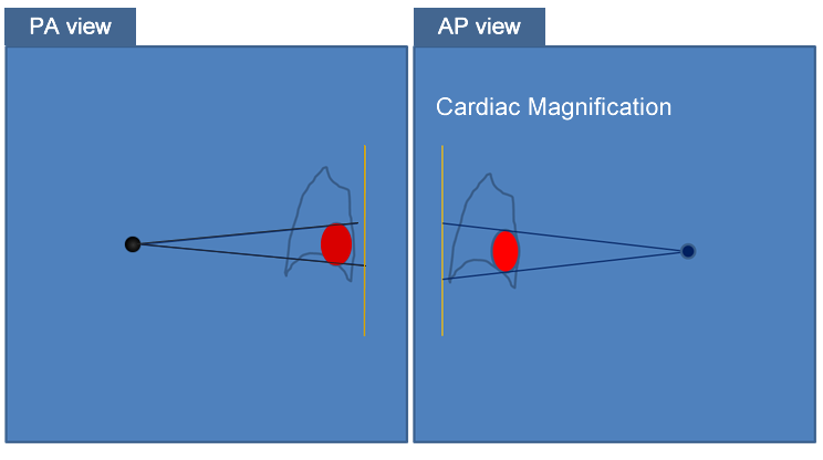
Here are two examples of a an PA and a AP film with the heart appearing much larger on the AP view just due to the way the Xray was taken.