3] Assessment of the Film
3.3] Hila
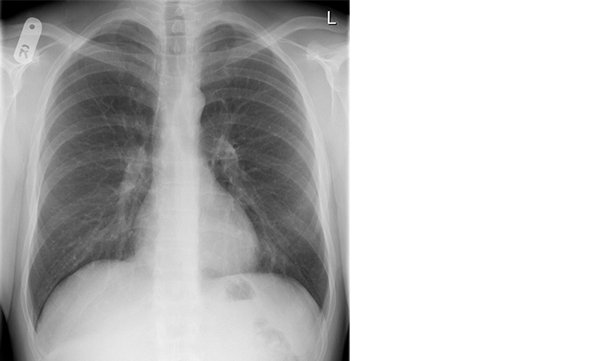
The hila comprise the lung roots which consist of the major bronchi, pulmonary arteries, veins entering and leaving the lungs as well as lymph nodes.
Hila Anatomy
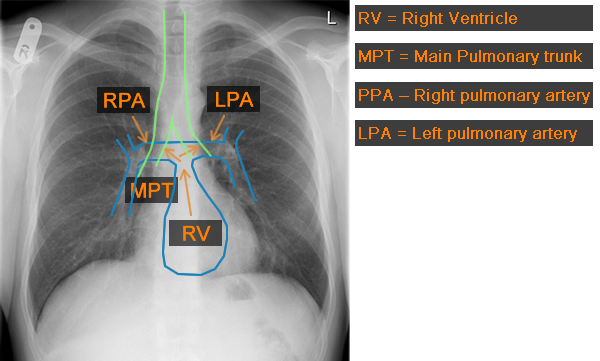
The hila comprise predominantly of the pulmonary arteries. The main vessel arising from the right ventricle is the main pulmonary trunk. The trunk leads into the right and left main pulmonary arteries. The left pulmonary artery passes over and behind the left main bronchus and therefore never lies above the right pulmonary artery. The right pulmonary artery lies in front of the bronchus but does not pass over the bronchus.

Hilar point
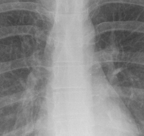
The hilar point which is often quoted is the point at which the upper lobe pulmonary veins meet the lower lobe pulmonary arteries. It should be a “v” shape and becomes distorted in the presence of enlarged lymph nodes.
Hila position
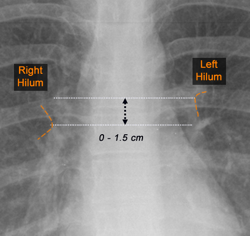
The position of the hilar are very important. The left hilum should always lie either at the same level or higher than the right as the left pulmonary artery hooks over the left main bronchus. The left hilum should NEVER BE BELOW the right! If the left hilum does lie lower it is a sign of left lower lobe volume loss.

A diagram to show both hila. The left hilum can be up to 1.5cm higher than the right hilum.
Hila size & density
When assessing the hila, it is important to look at there size, density and position. If they are increased in size and density it can be a sign of lymph node enlargement or pulmonary venous enlargement due to pulmonary hypertension. If the left hilum is pulled down it can be a sign of left lower lobe volume loss.


