3] Assessment of the Film
3.2] Lungs
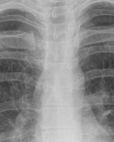
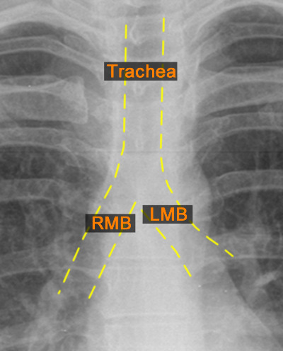
The anatomy of the upper airway is made up of the trachea which lies just to the right of the midline which splits into the right and left main bronchi at the carina which occurs roughly at the level of T4. It is airfilled and seen as a slightly blacker tubular structure on the Xray. The right main bronchus is steeper than the left. [For yellow and red labelling to appear on hovering]
Trachea & bronchi
The trachea can be seen as an air filled (black) tubular structure seen in the midline at the neck and extending down to the right. It splits into the right and left main bronchi at the carina.

Lung Parenchyma
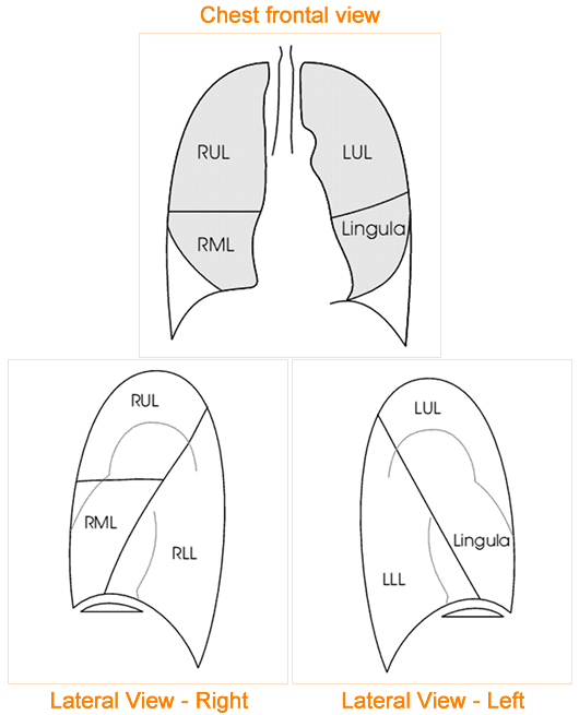
The lung Parenchyma consists of several lobes. The right lung comprises 3 lobes whereas the left lobe comprises only 2 lobes.
| Right 3 lobes | Left 2 lobes |
|---|---|
| Upper | Upper & Lingula |
| Middle | Lower |
| Lower |
The anatomy of the lungs can be appreciated by looking at frontal (AP) and lateral views. The right lung has both a horizontal (dividing the upper and middle lobes) and an oblique fissure (dividing the right upper and middle lobes form the lower lobe. The left lung however, only has an oblique fissure diving the upper and lingula from the lower lobe. (The lingula forms part of the left upper lobe).


