3] Assessment of the Film
3.5] Diaphragm
The diaphragm, the muscular layer separating the chest from the abdominal cavities. can also be assessed on a CXR. It appears dome shaped each side with the right is usually slightly higher due to the liver. The border with the heart produces the cardiophrenic recesses. The border with the chest wall produces the costophrenic recesses.
Pathology
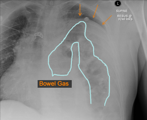
A number of different pathologies can occur to the diaphragm. It can become obscured if the lung adjacent to it becomes consolidated from infection, haemorrhage or oedema. Bowel loops containing air can rise through the diaphragm through a defect as in a hernia or free gas can be seen below it if a bowel viscus perforates. The diaphragm can also become raised if there is volume loss in the lungs or if underlying organs become enlarged.
Diaphragmatic Hernia
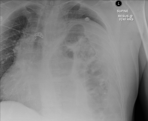
An example of a diaphragmatic hernia shown on CXR with the bowel loops seen within the left chest.
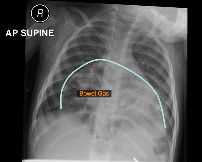
Morgagni Hhernia / Bowel Loops
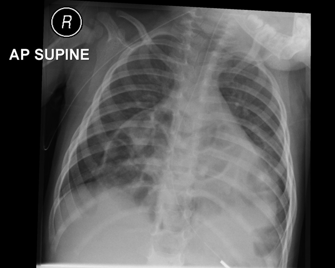
A further example of a Morgagni hernia or bowel loops passing through an anterior defect in the diaphragm. Bochdalek hernias occur when the defects are at the back.
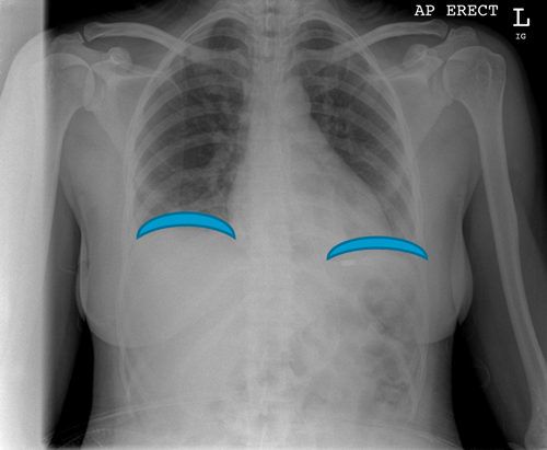
Typical Distribution of Free Gas
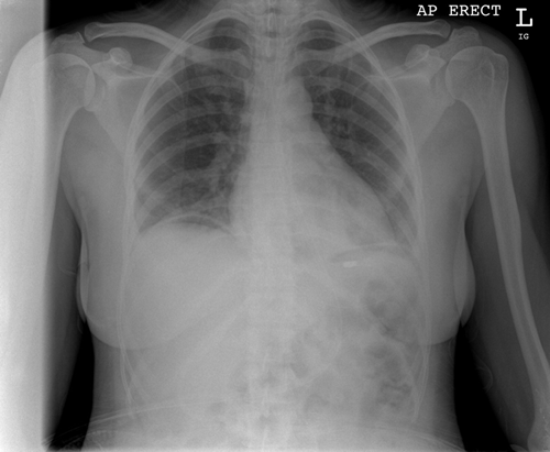
An example to show you the typical distribution of free gas under the diaphragm seen on a Erect CXR signalling likely perforation of an abdominal hollow viscus such as bowel.
