Right to Left Shunt
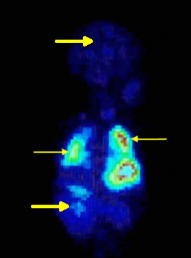
A right to left shunt with the tracer seen in
pulmonary (thin arrows pointing to the lungs) and systemic
(thick arrows pointing to the brain and kidneys) circulation
Right to left shunts can be estimated using various techniques.
Techniques for detecting right to left shunting:
- Colour Doppler echocardiography
- Hyperoxia test
- First pass radionuclide angiogram
- Radionclide labelled microsphere
Physiology of right to left shunts:
In a patient with a right to left shunt, following injection of labelled microspheres one notices that:
- Pulmonary capillary system is bypassed
- Particles enter the systemic circulation
- Particles are trapped in organs such as the brain and kidneys
Estimating right to left shunts using labelled microspheres involves carrying out a lung perfusion study. Regions of interest (ROI) are drawn around the anterior and posterior lungs and also the anterior and posterior whole body areas.
Shunt is estimated using the following formula: