Lung Perfusion Imaging in Children With Congenital Heart Disease
- Overview
- Examples
- Quantitation
Perfusion scintigraphy
This has a complementary role of assessing lung perfusion. This is more sensitive in in detecting:
- Reduced blood flow
- Small perfusion defects
Pulmonary arteriography
This gives an anatomical map of lung perfusion. This is more specific in identifying the cause of the perfusion abnormalities.
Complexity of cardiac heart disease
The indications for V/Q imaging in children are:
- Quantitation of relative perfusion
- Assessment of perfusion abnormalities
- Assessment of perfusion prior to surgery
- Assessment of lung perfusion post surgery/stent implantation/balloon angioplasty
- Assessment of right to left shunt
- Follow up studies
Clinical indications are:
- Pulmonary artery stenosis (PAS): branch pulmonary artery stenosis, a patent Blalock-Taussig shunt
- Pulmonary hypoplasia
- Pulmonary hypertension
- Suspected pulmonary embolus: patients who had cavopulmonary anastomosis
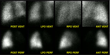
The following images illustrate changes in lung perfusion before and after intervention.
Click on each title to see a description.
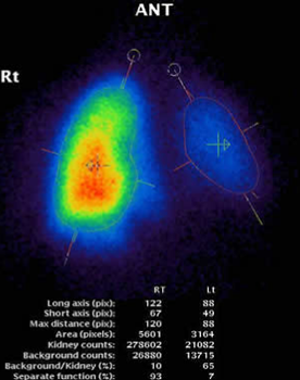
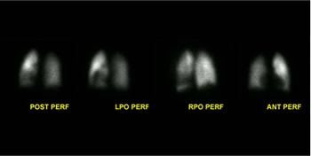
The image gives a split lung perfusion in a patient with left main pulmonary artery stenosis prior to surgery. The left lung is seen to contribute to 7% of total lung perfusion.
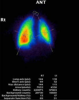
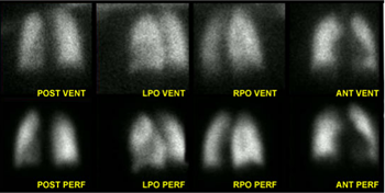
The above image gives the improved split lung perfusion in a patient with left main pulmonary artery stenosis post surgery. The left lung is now seen to contribute to 31% of total lung perfusion.
Consideration should be given to the following aspects:
- Careful choice of injection site
- Knowledge of the patient’s history and complex anatomy of cardiac malformations
- Knowledge of previous surgery
- Comparison with relevant angiograms and previous lung perfusion studies