-
Learning Units
- Unit 1: Sudden Loss of Vision
- Unit 2: Visual field defects, double vision & optic disc swelling
- Unit 3: Pupil abnormalities, Facial nerve palsy & Ptosis
- Unit 4: Refractive Error
- Unit 5: Children & Squint
- Unit 6: Differential diagnosis of blurred vision
- Unit 7: Gradual Loss of Vision
- Unit 8: Eye Trauma
- Unit 9: Red Eye
- Unit 10: Systemic Disease
- Useful Links
- KEATS
- KCL website
- Contact Us
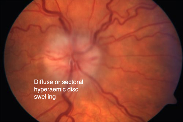
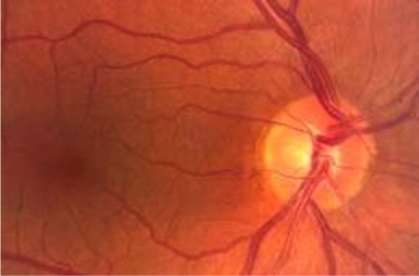
3: Optic Disc Swelling
Causes of Disc Swelling
Unilateral
- Central retinal vein occlusion
- Non arteritic anterior ischemic optic neuropathy
- Arteritic anterior ischemic optic neuropathy . This is a feature of giant cell arteritis.
- Papillitis
- Neuroretinitis
Bilateral
- Intracranial mass causing raised intra cranial pressure resulting in papilloedema
- Malignant hypertension
- Optic disc drusen produces pseudo papilloedema
Optic disc drusen
Image credit: retinagallery.com
Central Retinal Vein Occlusion
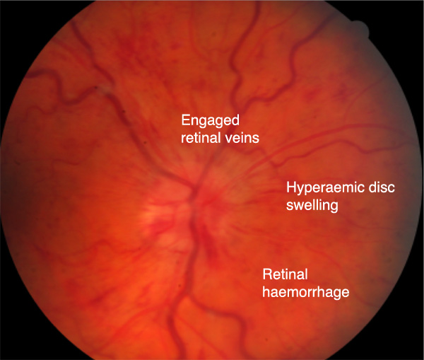 Source: Adapted from an image uploaded to retinagallery.com by Steven Cohen
- Common
- Usually unilateral
- Can cause severe visual impairment
Non-arteritic anterior Ischemic Optic Neuropathy
Non-arteritic anterior ischemic optic neuropathy (AION) is caused by occlusion of the short posterior ciliary arteries. This results in infarction of the optic nerve head.
Symptoms
- Painless monocular sudden loss of vision
Signs
- VA can vary from slightly to severely reduced
- A sector of the optic nerve may be swollen
- Splinter flame shaped haemorrhages around disc
- Occasionally macular star
Arteritic anterior ischemic optic neuropathy
- Sudden total monocular loss of vision
- May have been preceded by transient episodes of vision loss
- Ophthalmic emergency, other eye can develop loss within hours
- Age usually >50 Years
Symptoms
- Headache
- Scalp tenderness
- Loss of appetite
- Jaw Claudication
- Weight loss
Signs
- Visual acuity worse than count fingers
- Usually non pulsatile tender temporal artery
- Swollen optic disc & RAPD
- Flame haemorrhages & Cotton wool spots indicating nerve fibre death
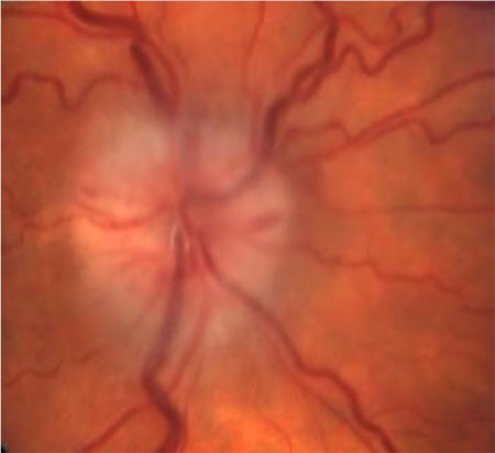
Pale swollen optic disc
Source: Adapted from an image uploaded to retinagallery.com by Steven Cohen
Relative afferent pupillary defect
A relative afferent pupillary defect (RAPD) is seen on examination by shining light in one eye, then the other.
If the left eye has an RAPD it will show little constriction of the pupil when a light is shone into that eye, but when the light is shone into the right eye, the left constricts.
When swinging back to the left eye it paradoxically dilates, as it is not as responsive to stimulation (as the optic nerve is damaged).
Papilloedema
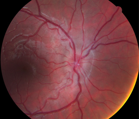
Source: Adapted from an image uploaded to retinagallery.com by Steven Cohen
Papilloedema refers to a swollen optic nerve head secondary to raised intracranial pressure.
Usually bilateral.
Malignant hypertension
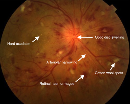
Source: Adapted from an image uploaded to retinagallery.com by Lucy James, COA
Malignant Hypertension
- Caused by accelerated raised blood pressure
- Clinically seen as Grade 4 vascular hypertension
Optic Disc Drusen

Image credit: retinagallery.com
Drusen (calcified deposits) can occur at the optic nerve head or deeper.
When located posterior to the disc they can give the appearance of a swollen optic nerve.
Can cause a visual field defect but usually asymptomatic.
No treatment is available.
© Faculty of Life Sciences & Medicine, Faculty Education Services, Digital Education Team. King's College London.
This module was prepared by C. Petrarca and T. Jackson and they retain the intellectual property rights in relation to its contents. The contents of this module must not be reproduced, or used outside King's, without their permission.

