Opthalmology
Learning Units
Unit 1: Sudden Loss of Vision
Unit 2: Visual field defects, double vision & optic disc swelling
Unit 3: Pupil abnormalities, Facial nerve palsy & Ptosis
Unit 4: Refractive Error
Unit 5: Children & Squint
Unit 6: Differential diagnosis of blurred vision
Unit 7: Gradual Loss of Vision
Unit 8: Eye Trauma
Unit 9: Red Eye
Unit 10: Systemic Disease
Useful Links
KEATS
KCL website
Contact Us
Unit 10: Systemic Disease
Introduction
1: Diabetic Retinopathy
Presentations
Prognosis
Aetiology
Signs
Differential diagnosis
History
Examination
Management 1
Management 2
Classification
2: Thyroid Eye Disease
Introduction
NOSPECS
Symptoms
Signs
Investigations
Management
3: Multiple Sclerosis
Introduction
Symptoms
Signs
History
Examination
4: HIV
Introduction
History & Examination
Signs
5: Hypertensive Retinopathy
Introduction
Symptoms
Signs
Classification
History & Examination
Differential diagnosis
6: Herpes Zoster
Introduction
Symptoms
Signs
Treatment
Self Assessment
5: Hypertensive retinopathy
Signs
Signs
×
Signs
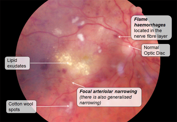
Signs of hypertensive retinopathy
Source:
of hypertensive retinopathy:
Focal arteriolar narrowing
Venous nipping at arteriovenous crossings
Copper and silver wiring
Cotton wool spots from small areas of retinal ischemia and swelling
Lipid (hard) exudates are yellow white intraretinal lipid collections from vascular leakage. At the macula they may resemble a star
Flame haemorrhages in the retinal nerve fibre layer
Macular oedema
Macro aneurysms seen as well defined red dots (sometimes with lipid exudates)
Optic disc swelling caused by local ischemia
«
Top
»